
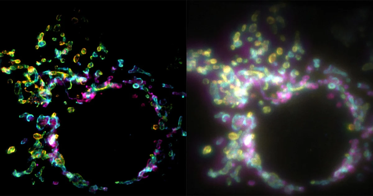
A new fluorescence microscopy technique, termed Spatial Polarization-Induced Fluorescence Fluctuation Imaging (SPIFFI), promises to revolutionize cellular biology by generating super-resolution images from a single exposure, offering scientists an extraordinary window into the dynamic inner workings of living cells. This innovative method directly addresses long-standing challenges associated with conventional super-resolution techniques, particularly their limitations in capturing rapid cellular processes due to reliance on multi-frame data acquisition. The research, spearheaded by Wei Guo, Lely Feletti, and Aleksandra Radenovic, and recently published in the prestigious journal Nature, marks a significant leap forward in our ability to observe biological phenomena at the nanoscale.
The Unseen World: Confronting the Diffraction Limit
For centuries, the fundamental barrier to observing the microscopic world with optical instruments has been the diffraction limit, a physical constraint articulated by Ernst Abbe in 1873. Abbe’s law dictates that an optical microscope cannot resolve objects closer than approximately half the wavelength of the light used to image them. For visible light, this typically means a resolution limit of around 200-250 nanometers (nm). While this resolution is sufficient to visualize larger cellular structures like nuclei or mitochondria, it renders many crucial biological components—such as individual proteins, molecular complexes, and synaptic vesicles, which are often tens of nanometers in size—indistinguishable. This optical blur has long obscured the intricate dance of molecules and organelles that define life at its most fundamental level.
The Dawn of Super-Resolution Microscopy: A Nobel-Winning Endeavor
The quest to overcome the diffraction limit has driven immense innovation in microscopy. The early 21st century witnessed the emergence of groundbreaking techniques that collectively broke Abbe’s barrier, earning Eric Betzig, Stefan Hell, and William Moerner the Nobel Prize in Chemistry in 2014 for "the development of super-resolved fluorescence microscopy." These methods fundamentally reshaped our understanding of cellular architecture and function.
One such technique, Stimulated Emission Depletion (STED) microscopy, developed by Stefan Hell, uses a donut-shaped "depletion" laser beam to switch off fluorescence at the periphery of a desired spot, effectively shrinking the fluorescent area and achieving higher resolution. Another category, Single-Molecule Localization Microscopy (SMLM), which includes Photoactivated Localization Microscopy (PALM) and Stochastic Optical Reconstruction Microscopy (STORM), pioneered by Betzig and Moerner, relies on switching individual fluorescent molecules on and off stochastically. By precisely localizing the centers of these isolated emitters over many frames, a super-resolved image can be computationally reconstructed. Structured Illumination Microscopy (SIM) is yet another approach that uses patterned light to generate moiré fringes, which carry high-resolution information that can be extracted computationally.
Limitations of Current Super-Resolution Techniques for Live-Cell Dynamics
Despite their transformative power, these established super-resolution techniques present significant challenges, especially when attempting to image living biological systems. The primary hurdle lies in their reliance on multi-frame data acquisition.
- Speed: STED, PALM, and STORM often require hundreds, if not thousands, of individual images to build a single super-resolved frame. This extensive data collection translates into acquisition times ranging from seconds to minutes for a single image, making it exceedingly difficult to capture fast-moving biological processes that occur on millisecond timescales, such as protein trafficking, membrane dynamics, or the rapid structural changes during cell division.
- Phototoxicity and Photobleaching: The prolonged and intense laser illumination necessary for multi-frame imaging significantly increases phototoxicity, damaging delicate living cells and altering their natural behavior. Moreover, fluorophores (the fluorescent molecules) can undergo photobleaching, losing their ability to emit light, which limits the duration of observation and the total number of images that can be acquired.
- Complexity and Cost: Many super-resolution setups are technically complex, requiring specialized lasers, optics, and sophisticated computational reconstruction algorithms. This often translates to high equipment costs and steep learning curves for researchers, limiting their widespread adoption in all research labs.
- Motion Artifacts: For dynamic live cells, movement during the long acquisition times of multi-frame methods can introduce motion artifacts, blurring the reconstructed super-resolved images and making accurate interpretation challenging. Correcting these artifacts adds another layer of computational complexity.
These limitations have, to a degree, confined super-resolution microscopy to fixed, non-living samples, leaving a critical gap in our ability to observe the real-time, nanoscale events that underpin life.
SPIFFI: A Novel Approach to Seeing the Unseen
Enter SPIFFI, Spatial Polarization-Induced Fluorescence Fluctuation Imaging, a technique developed by researchers at EPFL’s Laboratory of Nanoscale Biology (LBEN), led by Professor Aleksandra Radenovic. This innovative method directly confronts the speed and phototoxicity issues by achieving super-resolution from a single exposure. The core innovation of SPIFFI lies in its clever utilization of the inherent polarization properties of fluorescent light, a characteristic often overlooked or underutilized in conventional fluorescence microscopy.
"Essentially, previous approaches used temporal information to resolve spatial resolution, but this doesn’t work very well on living cells," explains Wei Guo, the first author and an LBEN PhD student. "With SPIFFI, every frame is super-resolved, meaning we can now produce super-resolution videos of live cells." This statement encapsulates the paradigm shift SPIFFI represents: moving from static snapshots to dynamic, high-resolution movies of cellular life.
How SPIFFI Works: Harnessing Polarization for Enhanced Detail
The mechanism behind SPIFFI leverages a fundamental property of fluorescent molecules: when excited by light, they emit polarized light. This means that the light waves emitted by a fluorescent molecule do not oscillate randomly in all directions but rather preferentially in certain orientations, dictated by the molecule’s spatial alignment at the moment of emission. Different molecules, or the same molecule in different orientations within a structure, will emit light with distinct polarization signatures.
SPIFFI ingeniously exploits this phenomenon. Instead of capturing a single, integrated image, the system splits the fluorescent light emitted from the sample into four distinct polarization-sensitive channels. Each channel records a slightly different aspect of the emitted light’s polarization state. By comparing the images obtained from these four channels, the technique can mathematically recover previously hidden structural details that would otherwise be blurred by the diffraction limit in a conventional widefield microscope. This multi-channel polarization analysis allows for the extraction of spatial information that is typically lost in a single, unpolarized image.
The principle is akin to how our brains infer depth from two slightly different images captured by our eyes. SPIFFI uses differences in polarization patterns across multiple detection channels to infer structural information at a sub-diffraction limit. This spatial encoding of information, rather than temporal encoding (as in multi-frame methods), is what allows SPIFFI to achieve super-resolution in a single shot.
Quantifiable Breakthroughs: Resolution and Speed
The experimental results validating SPIFFI’s efficacy are compelling. The researchers demonstrated that the technique can improve image resolution by a factor of two in a single exposure, effectively halving the observable feature size compared to traditional widefield microscopy. This means that structures around 160-170 nanometers in size can be distinctly resolved, a significant improvement over the typical 250nm diffraction limit.
Furthermore, the research indicates that when SPIFFI images are seamlessly integrated with existing fluctuation-based methods for post-processing—a technique that can be applied after the initial single-exposure acquisition—the resolution can be further enhanced to approximately 80 nanometers. This combined approach offers a powerful pathway to even finer detail without sacrificing the initial speed advantage.
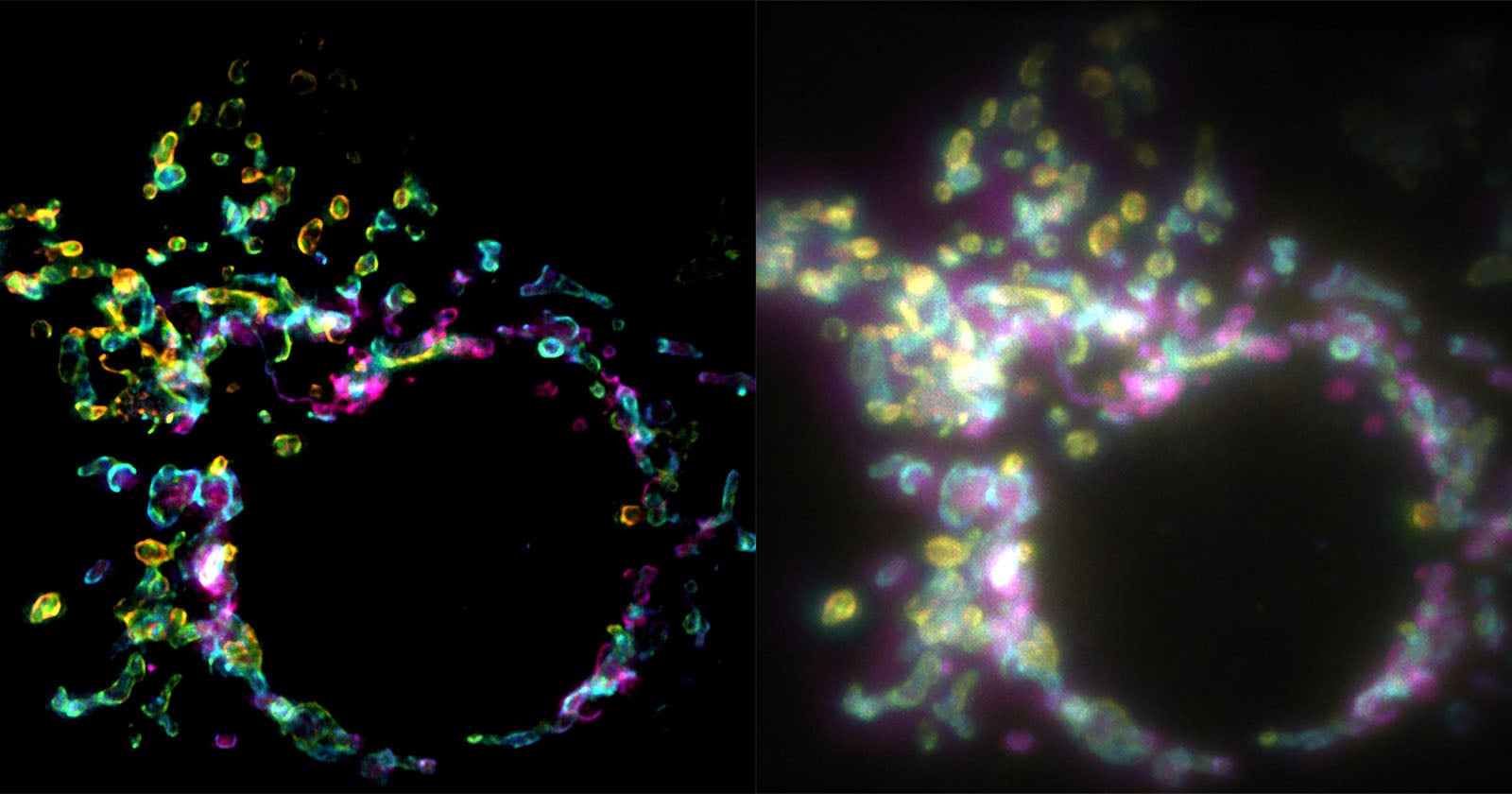
The ability to achieve super-resolution from a single frame means that SPIFFI can capture dynamic events that unfold rapidly. The researchers have successfully used SPIFFI to sharply image fast-moving biological phenomena, such as cellular splitting and fusion, processes that involve rapid morphological changes and molecular reorganizations. This capability fundamentally transforms the scope of biological questions that can be addressed, moving beyond static structural analysis to dynamic functional observation.
Enabling Live-Cell Dynamics: A New Era for Biological Observation
The most profound implication of SPIFFI’s single-exposure super-resolution capability is its potential to unlock the secrets of live-cell dynamics. Living cells are incredibly active environments; organelles constantly move, proteins are synthesized and transported, membranes fuse and divide, and signaling pathways activate and deactivate within milliseconds. Traditional super-resolution techniques, with their inherent slowness and phototoxicity, have largely been confined to providing snapshots of these processes, often requiring cells to be fixed or subjected to conditions that compromise their natural state.
SPIFFI changes this paradigm. "SPIFFI can capture fast-moving processes within cells, while enabling high-throughput, multi-dimensional imaging beyond the limits of conventional microscopes," states Professor Radenovic, emphasizing the technique’s capacity for both speed and comprehensive data acquisition. The ability to generate super-resolved videos of live cells means that scientists can now track the precise movements of individual molecules or nanoscale structures as they participate in crucial cellular functions. This opens avenues for studying phenomena such as:
- Viral entry and budding: Observing how viruses interact with and invade host cells at the molecular level.
- Synaptic transmission: Visualizing the rapid release and uptake of neurotransmitters in neuronal synapses.
- Mitochondrial dynamics: Tracking the fusion and fission events of mitochondria, vital for cellular energy regulation.
- Membrane trafficking: Following the pathways of vesicles and cargo within the cell.
- Cytoskeletal remodeling: Witnessing the dynamic assembly and disassembly of the cell’s internal scaffolding.
By providing dynamic, high-resolution insights, SPIFFI is poised to dramatically accelerate our understanding of disease mechanisms, drug interactions, and fundamental biological processes.
Accessibility and Integration: A Practical Innovation
Beyond its technical prowess, SPIFFI offers a significant practical advantage: its optical hardware can be implemented into existing fluorescence microscopes. This "retrofitting" capability is a crucial factor for its widespread adoption in the scientific community. Unlike many cutting-edge microscopy systems that demand entirely new, dedicated, and often astronomically expensive setups, SPIFFI can potentially upgrade standard laboratory microscopes. This significantly lowers the barrier to entry for research groups, making super-resolution imaging more accessible to a broader range of scientists.
The ability to integrate SPIFFI into current infrastructure implies a more cost-effective pathway to advanced imaging. It reduces the need for laboratories to invest in entirely new microscopy platforms, instead allowing them to leverage their existing equipment with a relatively straightforward addition. This pragmatic approach is likely to hasten its uptake across various biological disciplines.
The researchers are not resting on their laurels; they are actively working to make SPIFFI even more compact. Further miniaturization of the optical components will not only improve its usability but also potentially allow for integration into an even wider array of microscopy platforms, expanding its reach and impact.
Broader Implications for Biological Research and Beyond
The implications of SPIFFI extend across numerous fields of biological and medical research. In neuroscience, understanding the nanoscale architecture and dynamics of synapses is crucial for unraveling the mechanisms of learning, memory, and neurological disorders. SPIFFI could offer unprecedented views of neurotransmitter release, receptor dynamics, and structural plasticity. In immunology, tracking the interactions between immune cells and pathogens, or the formation of immunological synapses, could lead to new strategies for vaccine development and immunotherapy.
For cancer research, observing the dynamic changes in cellular morphology, membrane receptors, and cytoskeletal components during metastasis or in response to therapeutic agents could provide critical insights into disease progression and treatment efficacy. In developmental biology, the ability to image rapidly evolving cellular structures during embryogenesis could shed light on fundamental processes of tissue formation and organ development.
Beyond basic science, SPIFFI’s capabilities could also accelerate drug discovery. By enabling real-time, high-resolution observation of drug-target interactions within living cells, researchers can gain a more precise understanding of how potential therapeutics exert their effects, leading to more efficient screening and development of new medicines. In diagnostics, future iterations of such technology could potentially aid in the early detection of disease by identifying subtle nanoscale cellular abnormalities that are currently invisible.
The Road Ahead: Future Developments and Challenges
While SPIFFI represents a monumental step forward, the journey of innovation continues. The researchers’ immediate goal to make the system more compact will undoubtedly enhance its practicality and integration capabilities. Future developments might also focus on expanding its multi-color imaging capabilities, allowing for the simultaneous tracking of several different molecular species at super-resolution. Further refinements in computational algorithms could also push the resolution limits even lower, potentially approaching molecular dimensions without sacrificing speed.
Potential challenges include ensuring robust performance across a wide range of biological samples and experimental conditions, standardizing protocols for data acquisition and analysis, and continuing to mitigate any residual phototoxicity or photobleaching effects as imaging duration or intensity increases. However, the fundamental breakthrough of single-exposure super-resolution using polarization offers a robust foundation for overcoming these hurdles.
In conclusion, SPIFFI is not merely an incremental improvement; it is a transformative technology poised to open up entirely new avenues of inquiry in cell biology. By offering a rapid, high-resolution, and relatively accessible means to peer into the living, moving cell, it empowers scientists to finally observe the dynamic nanoscale processes that are the very essence of life, promising a future where the previously unseen becomes vividly clear.